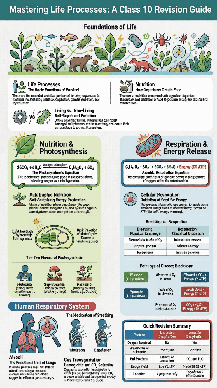
Life Processes Class 10 Biology Notes: Life processes are the basic activities that keep living organisms alive and functioning. In Life Processes Class 10 Biology Notes, students learn how the human body and other living organisms perform essential functions such as nutrition, respiration, transportation, and excretion. These processes help organisms obtain energy, grow, repair damaged cells, and maintain proper body balance. Understanding these concepts is very important for students preparing for the CBSE Class 10 Science exam.
These notes of life processes class 10 are designed to explain important biological concepts in simple and clear language so that students can easily understand them. The chapter mainly focuses on how living organisms take in food, break it down to release energy, transport nutrients and oxygen through the body, and remove waste materials. Along with explanations, students can also practice life processes class 10 questions and answers to test their understanding and improve exam preparation.
In these life processes class 10 science notes, diagrams, examples, and short explanations help students quickly revise key topics. Many students also look for life processes class 10 biology notes pdf download so that they can study anytime and revise important concepts before exams.
Overall, this chapter builds a strong foundation in biology by helping students understand how life is sustained through different biological systems. Some topics may look a bit complex at first, but with proper notes and regular practice it becomes easier to understand.
What Are Life Processes?
Life processes are the essential biological functions performed by living organisms to sustain their existence, maintain homeostasis, and ensure survival. Every living cell from a single-celled Amoeba to the trillions of cells in the human body depends on these processes.
CBSE Class 10 Biology Chapter 5 Life Processes & Nutrition Revision Notes PDF
Fill the form to download this PDF
The Seven Characteristics of Living Things
| Characteristic | Description |
|---|---|
| Movement (Locomotion) | Living things can move by themselves |
| Nutrition | Living things need food, air, and water |
| Growth | Living things can grow and develop |
| Control & Coordination | Living things respond to stimuli |
| Respiration | Living things release energy from food |
| Excretion | Living things remove metabolic waste |
| Reproduction | Living things can produce offspring |
Difference between Living vs. Non-Living Things
| Living Things | Non-Living Things |
|---|---|
| Have self-built organisation beyond individual level | Organisation is imposed and limited |
| Obtain simple molecules and convert them into complex protoplasmic constituents | No such activity seen |
| Perform growth, development, and differentiation | No such activity occurs |
| Have the property of self-repair | Repairs can only be done by external agencies |
| Have a definite life span | No definite life span |
| Reproduce and pass genes to next generations | Multiplication is imposed |
| Have the ability to evolve over time | Cannot evolve |
Note: The distinction between living and non-living lies in the concept of self-organisation and self-regulation living things maintain and perpetuate themselves through internally-driven biochemical processes.
Nutrition
Nutrition is the complete process by which an organism obtains food, and involves ingestion, digestion, absorption, assimilation, and egestion. A nutrient is any substance that nourishes a living being.
Functions of Food
Food serves multiple vital roles in the body:
- Provides energy to carry out work
- Helps maintain body temperature
- Supplies materials necessary for growth and reproduction
- Repairs damaged cells and tissues
- Assists in the removal of waste materials
- Maintains water balance in the body
Remember: Carbohydrates and fats primarily provide energy, while proteins and mineral salts are used for biosynthesis of body components (skin, blood, etc.).
Modes of Nutrition
NUTRITION
- Autotrophic (Self-feeding)
- Photosynthesis (e.g., Green plants)
- Chemosynthesis (e.g., Certain bacteria)
- Heterotrophic (Feeding on other organisms)
- Holozoic (e.g., Most animals)
- Saprophytic (e.g., Fungi, certain bacteria)
- Parasitic (e.g., Cuscuta, Plasmodium)
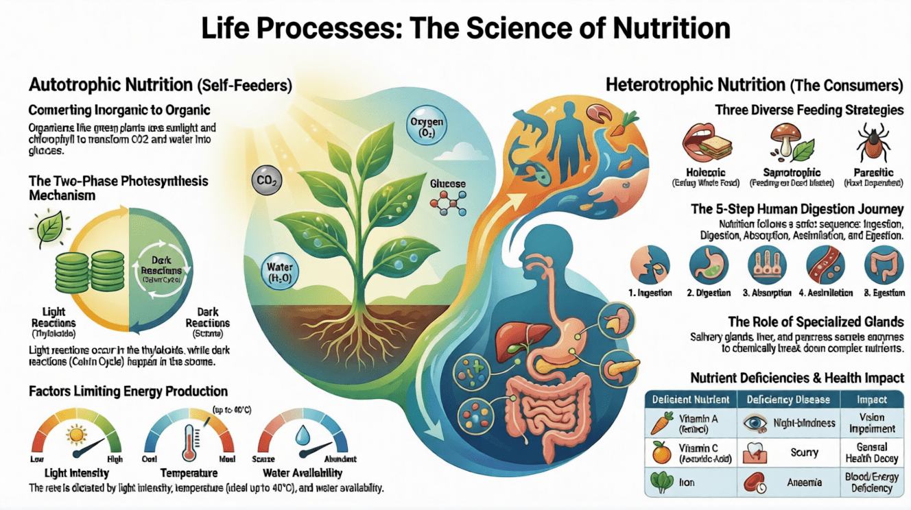
Autotrophic Nutrition & Photosynthesis
Autotrophic nutrition (Auto = self; trophic = food) is the mode in which organisms prepare their own food from inorganic raw materials.
There are two sub-types:
- Photoautotrophs: Use sunlight as the energy source (e.g., green plants)
- Chemoautotrophs: Use chemical energy (e.g., purple sulphur bacteria)
What is Photosynthesis?
Photosynthesis is the biochemical process by which green plants (and some bacteria) convert carbon dioxide and water into carbohydrates (glucose) using sunlight energy and chlorophyll, releasing oxygen as a by-product.
Overall Equation:
6CO₂ + 6H₂O ──(Chlorophyll / Sunlight) ──▶ C₆H₁₂O₆ + 6O₂
Where Does Photosynthesis Occur?
Photosynthesis takes place in chloroplasts, which are found mainly in the mesophyll cells of leaves. Chloroplasts are double-membrane structures containing:
- Stroma — the aqueous fluid (site of dark reactions)
- Grana — stacks of sac-like structures called thylakoids (site of light reactions)
- Stroma lamellae — connect thylakoids of different grana
Pigments involved: Chlorophyll (primary), Xanthophylls, and Carotene.
Mechanism of Photosynthesis
Photosynthesis occurs in two stages:
1. Light Reaction (Light-Dependent Reaction)
Occurs in the thylakoids of grana.
| Step | Process | Outcome |
|---|---|---|
| Step 1 | Excitation of chlorophyll by photons | Light energy → Chemical energy |
| Step 2 | Phosphorylation | ADP + Pi + Energy → ATP |
| Step 3 | Photolysis of water | H₂O → 2H⁺ + O₂ + 2e⁻ |
| Step 4 | Reduction of NADP | NADP + 2e⁻ + H⁺ → NADPH |
2. Dark Reaction (Light-Independent Reaction) — Calvin Cycle
Occurs in the stroma of chloroplasts. Does not require light directly.
- NADPH and ATP (from light reactions) are used to reduce CO₂ and produce sugar (C₆H₁₂O₆)
- Key intermediate: Ribulose-1,5-bisphosphate (RuBP)
- Stages: Carboxylation → Reduction → Regeneration
- Products eventually become: Glucose → Sucrose → Starch
Note: Desert plants (CAM plants) take up CO₂ at night and store it as malic acid, which is then used during the day.
Stomata - Gateway for Gas Exchange
Stomata are tiny pores on leaf surfaces guarded by two kidney-shaped guard cells.
- Turgid guard cells → stomata open (when water is sufficient)
- Flaccid guard cells → stomata close (when water is deficient)
- Desert plants keep stomata closed during daytime to prevent water loss
Factors Affecting Photosynthesis
| Factor | Effect |
|---|---|
| Light Intensity | Rate increases with light intensity up to a limit; very high intensity causes photo-oxidation (solarization) |
| Temperature | Rate increases up to 40°C; beyond this, enzymes are denatured and rate falls |
| CO₂ Concentration | Higher CO₂ increases rate (unless light or temperature is limiting); very high CO₂ is toxic to plants |
| Water | Water deficiency causes stomata to close, reducing CO₂ entry and thus photosynthesis |
Heterotrophic Nutrition
Heterotrophic nutrition (Hetero = different; trophic = nutrition) is the mode where organisms obtain energy by breaking down organic molecules prepared by other organisms.
Types of Heterotrophic Nutrition
1. Holozoic Nutrition Food in solid/liquid form is ingested into an alimentary canal for digestion and absorption.
- Herbivores: Feed only on plants (e.g., cow, goat, elephant)
- Carnivores: Feed on flesh of other animals (e.g., lion, tiger)
- Omnivores: Feed on both plants and animals (e.g., humans, dogs, crows)
2. Saprotrophic (Saprophytic) Nutrition Organisms live on dead/decaying organic matter, secrete digestive enzymes externally, and absorb the digested products.
Examples: Fungi, certain bacteria
3. Parasitic Nutrition An organism (parasite) derives food from another living organism (host) without killing it.
Examples: Cuscuta (dodder plant), Plasmodium, roundworms
Nutrition in Amoeba
Amoeba uses holozoic nutrition with intracellular digestion. The process involves five steps:
The Five Steps of Nutrition in Amoeba
1. Ingestion
- No fixed mouth; food intake occurs at any part of the body (usually the advancing end via pseudopodia)
- Process is called phagocytosis
- Food is engulfed into a food vacuole
2. Digestion
- Occurs inside food vacuoles
- Lysosomes (full of enzymes) fuse with food vacuoles
- Complex food molecules are broken down into simpler, diffusible forms
3. Absorption
- Soluble digested food diffuses from food vacuoles into the surrounding cytoplasm
4. Assimilation
- Absorbed food is synthesised into protoplasm by enzymes
- Excess food is stored as glycogen and fats
5. Egestion
- Undigested matter becomes denser and moves to the plasma membrane (plasmalemma)
- The plasmalemma ruptures at that point, ejecting the waste
- The plasmalemma is then repaired quickly
Nutrition in Human Beings
The human digestive system consists of:
- Alimentary canal (~9 metres long): Mouth → Buccal cavity → Oesophagus → Stomach → Small intestine → Large intestine → Anus
- Associated digestive glands: Salivary glands, Liver, Pancreas
Step-by-Step Journey of Food
The Mouth
Teeth types (Heterodont):
- Incisors — cutting
- Canines — tearing
- Premolars — grinding
- Molars — grinding
Dental formula (Permanent teeth): i²/₂, c¹/₁, pm²/₂, m³/₃ = 32 total teeth
Three pairs of salivary glands:
- Parotid glands — watery fluid rich in amylase
- Sub-mandibular glands — more viscous fluid
- Sublingual glands — thick, stringy saliva
Salivary amylase (Ptyalin) begins starch digestion → Maltose
Oesophagus
- ~25 cm long tube from pharynx to stomach
- Food moves by peristalsis (rhythmic contraction and relaxation of muscles)
- Oesophageal sphincter prevents regurgitation
Stomach
- J-shaped muscular bag below the diaphragm
- Divided into: Cardiac, Fundic, and Pyloric regions
- Pyloric sphincter controls food release into small intestine
Gastric gland cell types:
| Cell Type | Secretion | Function |
|---|---|---|
| Mucus cells | Mucus | Protects stomach lining |
| Chief cells | Pepsinogen (→ Pepsin via HCl) | Protein digestion |
| Parietal cells | Hydrochloric acid (HCl) | Activates pepsinogen; kills microbes |
Peptic Ulcer: Caused by imbalance in gastric acid secretion that erodes the mucosal lining of the stomach.
Small Intestine (~7 metres)
| Part | Length | Key Function |
|---|---|---|
| Duodenum | ~12 finger-breadths | Receives bile (from liver) and pancreatic juice |
| Jejunum | ~2 metres | Further digestion and absorption |
| Ileum | ~4 metres | Completes digestion; major absorption site |
- Inner wall has intestinal villi (finger-like projections) → greatly increase surface area for absorption (~8× body surface)
- Succus entericus (intestinal juice) from ileum glands completes carbohydrate and protein digestion
Liver and Pancreas
Liver (largest gland, ~1.5 kg):
- Secretes bile → emulsifies fats (breaks into small globules), creates alkaline medium
- Regulates blood sugar by storing excess glucose as glycogen
- Produces RBCs in embryo; removes excess amino acids via deamination
Pancreas (exocrine function):
- Secretes pancreatic juice containing: Amylase (starch), Trypsin (proteins), Lipase (fats)
Large Intestine
- Absorbs water and electrolytes
- Parts: Caecum → Colon → Rectum → Anus
- No nutrient digestion; secretes mucus
Digestive Enzymes
| Gland | Enzyme | Substrate | Product |
|---|---|---|---|
| Salivary gland | Salivary amylase | Starch | Maltose |
| Gastric glands | Pepsin | Proteins | Peptones |
| Gastric glands | Rennin | Casein (milk) | Paracasein |
| Liver | Bile (no enzyme) | Fats | Fat droplets |
| Pancreas | Pancreatic amylase | Starch/Glycogen | Maltose |
| Pancreas | Trypsin | Proteins | Peptides |
| Pancreas | Lipase | Triglycerides | Fatty acids |
| Intestinal gland | Maltase | Maltose | 2 Glucose |
| Intestinal gland | Sucrase | Sucrose | Glucose + Fructose |
| Intestinal gland | Lactase | Lactose | Glucose + Galactose |
Vitamin Deficiency Diseases
| Disease | Deficient Nutrient |
|---|---|
| Xerophthalmia / Night-blindness | Vitamin A (Retinol) |
| Rickets (children) / Osteomalacia (adults) | Vitamin D (Calciferol) |
| Scurvy | Vitamin C (Ascorbic acid) |
| Beri-beri | Vitamin B₁ (Thiamine) |
| Pellagra | Vitamin B₃ (Niacin) |
| Pernicious anaemia | Vitamin B₁₂ (Cyanocobalamine) |
| Bleeding disease | Vitamin K |
| Kwashiorkor / Marasmus | Protein / Protein + Calories |
Respiration
Respiration is the biological process by which cells use oxygen to break down carbohydrates and other nutrients, releasing energy and producing CO₂ and water as by-products.
Overall Equation (Aerobic):
C₆H₁₂O₆ + 6O₂ ──▶ 6CO₂ + 6H₂O + Energy (38 ATP)
Distinction: Breathing is an extracellular process (no enzymes; no ATP); Respiration is an intracellular process (enzymes involved; ATP produced).
Metabolic Terms
| Term | Definition |
|---|---|
| Anabolism | Constructive metabolism — smaller molecules unite to form larger ones (e.g., photosynthesis) |
| Catabolism | Destructive metabolism — breakdown of large molecules releasing energy (e.g., respiration) |
| Metabolism | The sum total of anabolism and catabolism in living beings |
| ATP | Adenosine Triphosphate — the universal energy currency of all living cells |
Types of Respiration
Aerobic Respiration
Requires oxygen produces maximum energy.
Pathway:
Glucose (C6H12O6)
↓
[Cytoplasm – Glycolysis – Absence of O2]
Pyruvate (3-carbon) + 2 ATP + Energy
↓
[Mitochondria – Krebs Cycle + ETC – Presence of O2]
CO2 + H2O + 36 more ATP
Total ATP = 38 molecules
Stages of Aerobic Respiration:
| Stage | Location | Process |
|---|---|---|
| Glycolysis | Cytoplasm | Glucose → Pyruvate (2 ATP) |
| Pyruvate oxidation | Mitochondria | Pyruvate → Acetyl CoA |
| Krebs Cycle | Mitochondria | Acetyl CoA drives hydrogen-generating reactions |
| Electron Transport Chain (ETC) | Mitochondria | Hydrogen → ATP + H₂O (36 ATP) |
Anaerobic Respiration (Fermentation)
Occurs without oxygen; incomplete breakdown; less energy produced.
| Organism | Product | Equation |
|---|---|---|
| Yeast (absence of O₂) | Ethanol + CO₂ | C₆H₁₂O₆ → 2C₂H₅OH + 2CO₂ + Energy |
| Muscle cells (lack of O₂) | Lactic acid | C₆H₁₂O₆ → 2CH₃CHOHCOOH + Energy |
Why do muscles cramp?
During intense exercise, oxygen supply is insufficient, and muscles switch to anaerobic respiration. The build-up of lactic acid causes muscular cramps.
Aerobic vs. Anaerobic Respiration
| Feature | Aerobic | Anaerobic |
|---|---|---|
| Oxygen required | Yes | No |
| Substrate breakdown | Complete | Incomplete |
| End products | CO₂ + H₂O | Alcohol/Lactic acid (+CO₂ in yeast) |
| Location | Cytoplasm + Mitochondria | Cytoplasm only |
| ATP produced | 38 molecules | 2 molecules |
Human Respiratory System
Respiratory Organs (in sequence)
Nostrils → Nasal Cavity → Nasopharynx → Larynx → Trachea → Bronchi → Bronchioles → Alveoli (Lungs)
| Organ | Feature |
|---|---|
| Nostrils & Nasal Cavity | Filters dust; normalises air temperature with mucus |
| Nasopharynx | Junction of nasal cavity and larynx; guarded by epiglottis |
| Larynx (Adam's apple) | Voice box; contains vocal cords; prevents food entry into trachea |
| Trachea | 16–20 C-shaped cartilaginous rings prevent collapse |
| Bronchi | Bilateral branching of trachea into two lungs |
| Alveoli | Actual sites of gas exchange; ~700 million in lungs; surrounded by capillaries |
| Pleura | Double membrane covering the lungs; intrapleural pressure drives breathing |
| Diaphragm | Dome-shaped muscle separating thoracic and abdominal cavities |
Mechanism of Breathing
Inspiration (Inhalation): Diaphragm contracts (flattens) → Thoracic volume increases → Pressure decreases → Air rushes in
Expiration (Exhalation): Diaphragm relaxes → Thoracic volume decreases → Pressure increases → Air is expelled
Residual Volume: Even after forcible expiration, ~1500 mL of air remains in the lungs, ensuring continuous O₂ absorption and CO₂ release.
Pulmonary Air Volumes
| Volume | Value | Description |
|---|---|---|
| Tidal Volume (TV) | ~500 mL | Normal breathing (in or out) |
| Inspiratory Reserve Volume (IRV) | ~2000–2500 mL | Extra air forcibly inhaled |
| Residual Volume (RV) | ~1500 mL | Air always remaining in lungs |
| Vital Capacity (VC) | ~3500–4500 mL | TV + IRV + Expiratory Reserve |
| Total Lung Capacity (TLC) | ~5000–6000 mL | VC + RV |
Transport of Gases
- Oxygen transport: Carried by haemoglobin in RBCs as oxyhaemoglobin (HbO₂)
- CO₂ transport: More soluble than O₂; mostly transported dissolved in blood as carbonic acid (H₂CO₃)
- Carbon monoxide (CO) danger: CO binds haemoglobin ~200× more strongly than O₂, forming stable carboxyhaemoglobin (HbCO), preventing O₂ transport → potentially fatal
Respiratory Disorders
| Disorder | Description |
|---|---|
| Bronchial Asthma | Allergic bronchial spasm; expiratory wheeze |
| Pneumonia | Alveoli fill with mucus due to acute infection |
| Emphysema | Abnormal distension of bronchioles/alveolar sacs; loss of elasticity |
| Bronchitis | Inflammation of the bronchi |
Transportation
Transportation is the life process by which substances synthesised or absorbed in one body part are carried to other parts where they are consumed.
Transportation in Humans
Humans have two circulatory systems:
Transportation in Humans
- Blood Vascular System
- Blood (Plasma ~55% + Corpuscles ~45%)
- Blood Vessels (Arteries, Veins, Capillaries)
- Heart
- Lymphatic System
- Lymph
- Lymph Vessels
- Lymph Nodes
Blood Composition
Blood = Plasma (~55%) + Corpuscles (~45%)
| Component | Description | Function |
|---|---|---|
| Plasma | 92% water + 8% solutes (proteins, salts, nutrients) | Transports food, hormones, CO₂, waste |
| RBCs (Erythrocytes) | Biconcave, denucleated; ~5–5.5 million/mm³; life span 120 days | Carry O₂ via haemoglobin |
| WBCs (Leucocytes) | ~6000–8000/mm³; nucleated | Immunity and defence |
| Platelets (Thrombocytes) | 1,50,000–3,50,000/mm³; non-nucleated fragments | Blood clotting |
Spleen is called the graveyard of RBCs — old erythrocytes are removed here.
Blood Vessels — Comparative Study
| Feature | Arteries | Veins | Capillaries |
|---|---|---|---|
| Direction | Away from heart | Towards heart | Arterioles to venules |
| Blood type | Oxygenated (except pulmonary artery) | Deoxygenated (except pulmonary vein) | Changes from oxy to deoxy |
| Blood pressure | High | Low | Extremely low |
| Blood flow | Rapid, with jerks | Smooth | Smooth |
| Lumen | Narrow | Wide | Very small |
| Semilunar valves | Absent | Present | Absent |
The Human Heart
The heart is a hollow, muscular, four-chambered organ lying between the lungs, above the diaphragm.
Structure
- 4 Chambers: Right atrium, Right ventricle, Left atrium, Left ventricle
- Auricles (Atria): Receiving chambers (thin walls)
- Ventricles: Pumping chambers (thick walls — especially left ventricle)
- Valves:
- Tricuspid valve — between right atrium and right ventricle
- Bicuspid (Mitral) valve — between left atrium and left ventricle
- Semilunar valves — at origins of aorta and pulmonary artery
Blood Flow Through the Heart
Blood Circulation Pathway
Body (deoxygenated blood)
↓ [Vena Cava]
Right Atrium → Right Ventricle
↓ [Pulmonary Artery]
Lungs (oxygenation)
↓ [Pulmonary Vein]
Left Atrium → Left Ventricle
↓ [Aorta]
Body (oxygenated blood)
Pulmonary circulation = Heart ↔ Lungs
Systemic circulation = Heart ↔ Rest of body
Cardiac Cycle (0.8 seconds)
| Phase | Duration |
|---|---|
| Atrial systole (contraction) | 0.1 sec |
| Ventricular systole (contraction) | 0.3 sec |
| Complete diastole (relaxation) | 0.4 sec |
- Heart rate: 70–72 beats/minute (adult male); 80/min (adult female)
- Heart sounds: "LUBB" (AV valves close) and "DUBB" (Semilunar valves close)
- Blood pressure: Normal = 120/80 mmHg (measured by sphygmomanometer)
- ECG: Electrocardiogram — graphic record of cardiac impulse conduction
SA Node and Impulse Conduction
Heart is myogenic (heartbeat is self-initiated by cardiac muscle):
SA Node (Pacemaker) → AV Node (Pacesetter) → Bundle of His → Purkinje Fibres
Heart Structure in Other Animals
| Animal | Chambers | Notes |
|---|---|---|
| Fish | 2 (1 auricle + 1 ventricle) | Blood oxygenated in gills |
| Amphibians | 3 (2 auricles + 1 ventricle) | Some mixing of blood tolerated |
| Reptiles | 3 + incomplete partition | Exception: Crocodile has 4 chambers |
| Birds & Mammals | 4 | Complete separation; high metabolic rate |
The Lymphatic System
- Lymph: Colourless fluid (tissue fluid) lacking RBCs, platelets, and plasma proteins; drains into blood circulatory system
- Lymph nodes: Filter lymph; rich in phagocytes and lymphocytes
- Flow direction: Unidirectional — from tissues to heart
- Key organs: Thymus and Spleen
Functions of Lymph:
- Transports fats from intestine to blood
- Returns metabolic substances (e.g., proteins) to blood
- Lymphocytes defend the body against infection
- Provides nutrition to areas where blood cannot reach
Transportation in Plants
Plants use a specialised vascular system for transport, consisting of:
Xylem — Water and Mineral Transport
Xylem tissue components: Vessels, Tracheids, Fibres, Parenchyma
The upward movement of water from roots to leaves = Ascent of Sap
Two key theories:
1. Root Pressure Theory:
- Roots absorb water and create osmotic pressure that pushes water upward
- Applicable in small herbs; more effective at night
2. Transpiration Pull & Cohesion-Tension Theory:
- Water evaporates from leaves (transpiration), creating a pull
- Cohesion of water molecules keeps the column continuous
- The main mechanism for tall trees; operative during daytime
Transpiration
Transpiration = loss of water as vapour from the aerial parts of a plant.
- ~80–90% through stomata
- ~9% through cuticle
- ~1% through lenticels
Functions of Transpiration:
- Drives ascent of sap (absorption and upward movement of water)
- Regulates leaf temperature (cooling effect via evaporation)
Potometer — instrument used to measure the rate of transpiration.
Phloem - Food Transport (Translocation)
Phloem tissue components: Sieve tubes, Companion cells, Phloem parenchyma, Phloem fibres
- Translocation: Transport of food (sucrose, amino acids) from leaves to other plant parts
- Can be bidirectional (up or down, depending on need)
Mechanism (Mass Flow Hypothesis — Munch, 1931):
- Sugar (sucrose) loaded into sieve tubes using ATP energy
- Osmotic pressure in sieve tubes increases
- Water enters sieve tubes by osmosis
- Material flows from high pressure (source — leaves) to low pressure (sink — roots/fruits)
Excretion
Excretion is the biological process of removal of harmful metabolic wastes from the body of an organism.
Types of Nitrogenous Waste
| Category | Waste Product | Toxicity | Example Organisms |
|---|---|---|---|
| Ammonotelic | Ammonia | Highly toxic; needs lots of water | Aquatic animals, Amoeba, Fishes |
| Ureotelic | Urea | Less toxic | Mammals (humans), Amphibians (frogs) |
| Uricotelic | Uric acid | Least toxic; excreted as solid | Birds, Reptiles, Insects |
Human Excretory System
Components: Pair of kidneys → Pair of ureters → Urinary bladder → Urethra
Structure of the Kidney
- Bean-shaped organs located on either side of the vertebral column
- Surrounded by renal capsule (fibrous connective tissue)
- Regions: Cortex (outer) → Medulla (inner) → Renal Pelvis (innermost) → Ureter
The Nephron — Functional Unit of the Kidney
Each kidney has ~1 million nephrons. Each nephron has 4 regions:
| Part | Location | Function |
|---|---|---|
| Bowman's Capsule + Glomerulus (Malpighian body) | Renal cortex | Ultra-filtration |
| Proximal Convoluted Tubule (PCT) | Renal cortex | Reabsorption of glucose, amino acids, salts, water |
| Loop of Henle | Renal medulla | Concentration of urine |
| Distal Convoluted Tubule (DCT) | Renal cortex | Tubular secretion; water regulation |
Three Processes of Urine Formation
1. Glomerular Ultrafiltration:
- Blood forced through glomerular capillaries under high pressure
- Water, urea, salts, glucose filtered out (large proteins stay in blood)
2. Tubular Reabsorption:
- Useful substances (glucose, amino acids, salts, water) reabsorbed back into blood from the renal tubule
3. Tubular Secretion:
- Excess potassium, hydrogen ions, creatinine, and drugs are secreted into the tubule from blood
Composition of Urine (Normal)
| Component | Percentage |
|---|---|
| Water | 95% |
| Urea | 2% |
| Uric acid | 0.3% |
| Creatinine | 0.1% |
| Sodium | 0.1% |
| Potassium | 0.6% |
Osmoregulation and ADH
- Osmoregulation: The kidney regulates water-salt balance in blood
- ADH (Antidiuretic Hormone) from the pituitary gland controls the permeability of DCT and collecting ducts
- More water in blood → Less ADH → Less reabsorption → More dilute urine
- Less water in blood → More ADH → More reabsorption → Concentrated urine
Accessory Excretory Organs
| Organ | Waste Excreted |
|---|---|
| Skin (sweat glands) | Water, salts, urea, lactic acid (as sweat) |
| Liver | Bile pigments (bilirubin, biliverdin) from haemoglobin breakdown |
| Lungs | CO₂, water vapour, aromatic substances |
| Large intestine | Heavy metals, toxins into faeces |
Artificial Kidney (Dialysis)
Used when both kidneys fail. Blood is taken from the radial artery, passed through semipermeable membrane tubes in a dialysis solution, and returned purified through the radial vein. Dialysis solution has same concentration as normal plasma.
Excretion in Plants
Plants lack a dedicated excretory system. They manage waste through:
- Diffusion/osmosis: Gaseous wastes (CO₂, O₂, water vapour) released through stomata and lenticels
- Cellular vacuoles: Store non-diffusible waste products
- Leaf shedding: Removes accumulated wastes stored in leaves
- Secretory structures: Tannins, essential oils, latex, gums, resins stored in bark, fruits, or leaves
- Aquatic plants: Diffuse metabolic wastes directly into surrounding water
Note: CO₂ produced during daytime respiration is directly used in photosynthesis so plants excrete oxygen as waste only during the day. At night, only CO₂ is excreted (from respiration alone).
Life Process Class 10 Science Solved Examples
Q. Write the balanced chemical equation for photosynthesis.
A.
6CO₂ + 6H₂O ──(Chlorophyll / Sunlight)──▶ C₆H₁₂O₆ + 6O₂
Q. Name the enzymes present in pancreatic juice.
A. Pancreatic juice contains three enzymes:
- Trypsin — digests proteins
- Pancreatic amylase — digests starch and glycogen
- Lipase — digests triglycerides into fatty acids and glycerol
Q. Define transpiration.
A. Transpiration is the loss of water in the form of water vapour through the stomatal pores of leaves (and also through cuticle and lenticels) in plants.
Q. Why is diffusion insufficient to meet the oxygen requirements of multicellular organisms like humans?
A. In a multicellular organism, all cells are not in direct contact with the surrounding environment. Simple diffusion cannot transport oxygen fast enough to reach every cell. A specialised circulatory system carrying oxygen via haemoglobin is required.
Q. In which form are carbohydrates stored in plants and in animals?
A.
- In plants: stored as starch
- In animals (and human body): stored as glycogen (in liver and muscles)
Q. What is the function of bile?
A. Bile emulsifies fats — it breaks large fat molecules into small globules, increasing the surface area for lipase enzyme action. It also creates an alkaline medium in the duodenum for pancreatic enzymes to work.
Q. How is oxygen transported in the human body?
A. Oxygen is transported in combination with haemoglobin (present in RBCs) as oxyhaemoglobin (HbO₂). Haemoglobin has a very high affinity for oxygen, making it efficient at picking up O₂ in the lungs and releasing it in tissues.
Q. Name the two processes involved in the mechanism of respiration (cellular level).
A. The two key processes are:
- Glycolysis (in cytoplasm)
- Krebs Cycle / Citric Acid Cycle (in mitochondria)
Q. Which organelle is the site of aerobic respiration?
A. Mitochondria often called the powerhouse of the cell because they produce ATP through aerobic respiration via the Krebs cycle and electron transport chain.
Q. How much energy (in ATP) is liberated by aerobic oxidation of one glucose molecule?
A. 38 ATP molecules are produced: 2 ATP from glycolysis + 2 ATP from Krebs cycle + 34 ATP from the electron transport chain.
Short Answer Type
Q. What is the role of hydrochloric acid (HCl) in the human digestive system?
A.
- HCl creates an acidic medium (pH ~2) in the stomach essential for protein digestion by pepsin
- It activates inactive pepsinogen into active pepsin
- It kills harmful microorganisms in food (disinfection)
- It denatures proteins, making them more susceptible to enzymatic digestion
Q. What are the various processes that take place in the duodenum?
A. Three key processes occur in the duodenum:
- Bile from the liver emulsifies fat molecules into small globules
- Pancreatic enzymes (trypsin, amylase) begin digesting proteins and starch respectively
- Bicarbonate ions secreted by the duodenal wall neutralise the acidic chyme, creating an alkaline medium for pancreatic enzymes to function optimally
Q. Why do ventricles have thicker walls than auricles?
A. Auricles (atria) only need to collect blood arriving from veins (low pressure) and push it a short distance into the ventricles. Ventricles must pump blood with enough force to reach distant organs — the right ventricle pushes blood to the lungs, and the left ventricle pumps blood to the entire body through the aorta. This requires thicker muscular walls to generate sufficient pressure.
Q. Why are valves present in veins but not in arteries?
A. Blood in arteries flows under high pressure generated directly by the heart's pumping action this pressure is enough to maintain unidirectional flow without valves. Blood in veins returns from body tissues under low pressure and must work against gravity in many parts of the body. Semilunar valves in veins prevent backflow and ensure blood moves only toward the heart.
Q. Write the overall equation of anaerobic respiration in yeast and in muscle cells.
A.
- In yeast:Glucose → Pyruvate → Ethanol + CO₂ + Energy (2 ATP)C₆H₁₂O₆ → 2C₂H₅OH + 2CO₂ + Energy
- In muscle cells:Glucose → Pyruvate → Lactic acid + Energy (2 ATP)C₆H₁₂O₆ → 2CH₃CHOHCOOH + Energy
Q. What advantage does a terrestrial organism have over an aquatic organism with regard to oxygen for respiration?
A. Aquatic organisms breathe oxygen dissolved in water, whose concentration is very low (~7 mg/L). Terrestrial organisms breathe atmospheric air where oxygen concentration is much higher (~21%). This means terrestrial organisms can obtain oxygen much more efficiently with less respiratory effort.
Q. Why is it necessary to separate oxygenated and deoxygenated blood in mammals and birds?
A. Birds and mammals are warm-blooded (homeothermic) — they maintain a constant high body temperature regardless of the environment. This requires significantly more energy and thus more efficient oxygen delivery. Complete separation of oxygenated and deoxygenated blood (via the 4-chambered heart) ensures that tissues always receive fully oxygenated blood at high pressure, maintaining the high metabolic rate needed to sustain body temperature.
Q. How do unicellular organisms remove metabolic waste products?
A. Unicellular organisms like Amoeba and Paramecium remove metabolic waste by simple diffusion from the body surface into the surrounding water. Some unicellular organisms also have specialised structures called contractile vacuoles that collect and expel excess water and dissolved wastes.
Q. How is the amount of urine produced regulated?
A. The amount of urine is regulated by ADH (Antidiuretic Hormone) secreted by the posterior lobe of the pituitary gland.
- When blood water level is low → More ADH is released → Increased permeability of DCT and collecting ducts → More water reabsorption → Less, concentrated urine
- When blood water level is high → Less ADH released → Less water reabsorption → More, dilute urine
Q. What happens to glucose that enters the nephron along with the filtrate during excretion in humans?
A. Glucose is a useful substance and is selectively reabsorbed by the blood capillaries (via active transport) as the glomerular filtrate flows through the proximal convoluted tubule (PCT). Under normal conditions, no glucose is present in the final urine. However, in diabetes mellitus, blood glucose is so high that the reabsorption capacity is exceeded, and glucose appears in the urine a condition called glycosuria.
Class 10 Science Life Process Notes Quick Revision
| Life Process | Key Organ (Humans) | Key Chemical | Energy |
|---|---|---|---|
| Nutrition (Digestion) | Alimentary canal + Glands | Enzymes (Amylase, Pepsin, Lipase) | — |
| Photosynthesis | Chloroplast (leaves) | Chlorophyll, ATP, NADPH | Sunlight → Chemical |
| Respiration | Mitochondria (cells) | ATP | Glucose → 38 ATP |
| Transportation | Heart, Blood, Xylem, Phloem | Haemoglobin, Sucrose | ATP (phloem) |
| Excretion | Kidneys, Lungs, Skin | Urea, CO₂ | — |
Exam Tips for Class 10 Students
- Memorise all chemical equations - photosynthesis, aerobic and anaerobic respiration equations are frequently tested.
- Draw and label diagrams - chloroplast structure, stomata, human digestive system, and alveoli diagrams carry significant marks.
- Understand enzyme-substrate specificity - know which enzyme acts on which substrate and in which organ.
- Compare aerobic vs. anaerobic respiration in a table format a very common exam question.
- Link structure to function - be able to explain WHY alveoli have a thin wall, WHY villi have large surface area, WHY trachea has cartilage rings.
- Remember vitamin deficiency diseases - create mnemonics to remember which vitamin deficiency causes which disease.