

You’re sitting in biology class, peering through a microscope at a drop of pond water. Suddenly, you see something incredible a tiny, slipper-shaped organism covered in what looks like hundreds of tiny hairs, moving gracefully through the water. Your teacher smiles and says, “That’s a paramecium, one of the most fascinating ciliated protozoans!”
For many students, this moment sparks curiosity about the microscopic world. Ciliated protozoans aren’t just another topic to memorize for exams—they’re living proof of nature’s incredible engineering. Understanding these organisms helps you grasp fundamental concepts in cell biology, evolution, and ecology. Whether you’re preparing for your biology test or simply curious about life’s diversity, this guide will make ciliated protozoans easy to understand and remember.

What Are Ciliated Protozoans?
Ciliated protozoans, or ciliates, are single-celled organisms belonging to the phylum Ciliophora. They’re called “ciliated” because their entire body surface is covered with tiny hair-like structures called cilia.
Key characteristics:
- Single-celled eukaryotic organisms
- Covered with cilia for movement and feeding
- Found in freshwater, marine, and moist soil environments
- Most are free-living and feed on bacteria or other microorganisms
Think of cilia like thousands of tiny oars that beat in coordinated waves. This coordinated beating allows the organism to swim, capture food, and interact with its environment all without a brain or nervous system!
How Ciliates Differ from All Other Protozoans
This is a crucial exam topic! Understanding how ciliates differ from all other protozoans helps you classify and identify these organisms correctly.
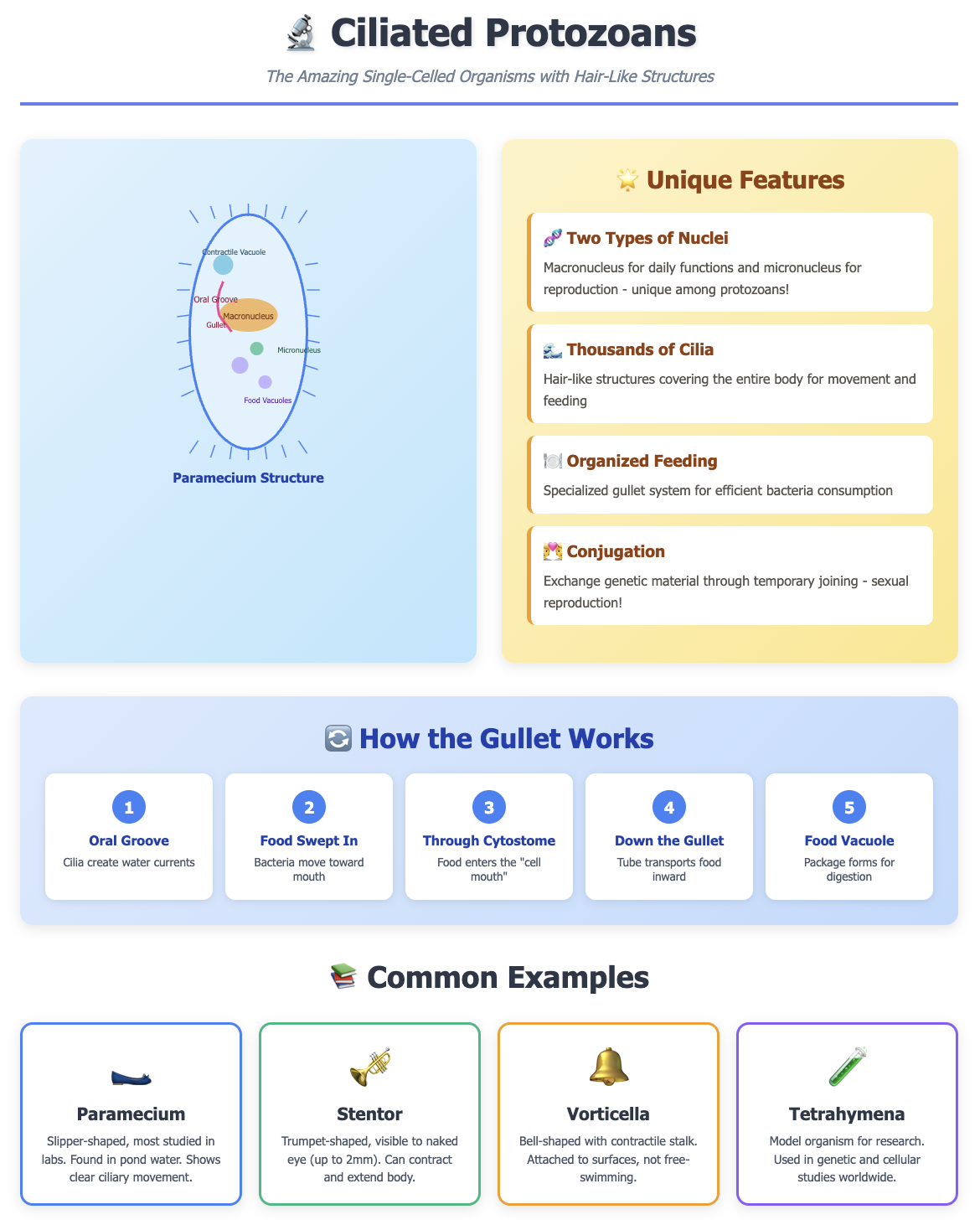
Nuclear Dimorphism
Ciliates are the only protozoans with two types of nuclei:
- Macronucleus: Controls daily cell functions (like eating and moving)
- Micronucleus: Stores genetic information for reproduction
Other protozoans have only one type of nucleus. This is like having both a work computer and a backup drive in one cell!
Complete Coverage of Cilia
While some other protozoans might have flagella (whip-like structures) or pseudopodia (temporary projections), ciliates are covered entirely or partially with cilia arranged in specific patterns. No other protozoan group shows this level of ciliary organization.
Complex Feeding Structures
Ciliates possess specialized structures like the cytostome (cell mouth) and gullet for ingestion—more organized than other protozoans’ feeding methods.

Sexual Reproduction Through Conjugation
Ciliates exchange genetic material through conjugation, where two organisms temporarily join and swap micronuclear material. This process is unique among protozoans.
Quick Comparison Table:
| Feature | Ciliates | Other Protozoans |
|---|---|---|
| Locomotion | Cilia | Flagella or pseudopodia |
| Nuclei | Two types (macro & micro) | One type |
| Feeding structure | Organized gullet system | Simple or no specialized structure |
| Reproduction | Conjugation + binary fission | Mainly binary fission |
Common Ciliated Protozoans Examples
Understanding ciliated protozoans examples makes abstract concepts concrete. Here are the most commonly studied in classrooms:
1. Paramecium (The Classic Example)
The superstar of biology labs! Paramecium looks like a tiny slipper and is found in freshwater ponds.
Student-friendly features:
- Slipper-shaped body
- Visible oral groove leading to gullet
- Easy to observe under school microscopes
- Shows clear ciliary movement
2. Stentor
Trumpet-shaped and often blue-green in color, Stentor can be seen with the naked eye (up to 2mm long).
Why students love it:
- Large enough to see without magnification
- Can contract and extend its body
- Often used in regeneration experiments
3. Vorticella
Bell-shaped ciliate attached to surfaces by a contractile stalk.
Classroom relevance:
- Demonstrates how not all ciliates swim freely
- Shows rapid stalk contraction when disturbed
- Common in aquarium samples
4. Tetrahymena
Used extensively in genetic and cellular research.
Academic importance:
- Model organism for cell biology
- Featured in advanced biology courses
- Won researchers Nobel Prizes for discoveries
5. Balantidium coli
The only ciliate known to cause disease in humans.
Medical relevance:
- Teaches about parasitic protozoans
- Important for microbiology students
- Shows practical importance of studying ciliates
Understanding the Gullet in Ciliated Protozoans
The gullet in ciliated protozoans is one of their most fascinating features. Many students struggle to understand this structure, but it’s simpler than you think.
What Is the Ciliated Protozoan Gullet?
The gullet (also called the cytopharynx) is a tubular structure that acts as the “throat” of the organism. It’s part of an organized feeding system that includes:
- Oral groove: A depression on the cell surface
- Cytostome: The “cell mouth” where food enters
- Gullet: The tube connecting the mouth to the interior
- Food vacuole formation site: Where food is packaged for digestion
How the Gullet Works (Step-by-Step)
Step 1: Cilia in the oral groove beat rhythmically, creating water currents Step 2: These currents sweep bacteria and food particles toward the cytostome Step 3: Food enters through the cytostome and moves down the gullet Step 4: At the end of the gullet, food is packaged into a food vacuole Step 5: The food vacuole detaches and moves through the cytoplasm for digestion
Real-life analogy: Think of the gullet system like a waterslide at a pool. The oral groove is the entrance where water flows in, the gullet is the slide itself, and the food vacuole formation is like landing in the pool at the bottom!
Why the Gullet Matters
The ciliated protozoans gullet shows evolutionary sophistication. Unlike amoebas that engulf food anywhere on their surface, ciliates have a designated feeding area making them more efficient hunters of bacteria.
Structure and Key Features
Understanding ciliate structure helps you ace diagram-based exam questions.
External Structures
Pellicle: A tough, flexible outer covering that maintains cell shape Cilia: Arranged in rows (called kineties) that beat in coordinated waves Trichocysts: Small dart-like structures for defense
Internal Structures
- Macronucleus: Large, kidney-shaped nucleus for metabolic control
- Micronucleus: Small, spherical nucleus for genetic storage
- Contractile vacuoles: Pump out excess water (crucial in freshwater species)
- Food vacuoles: Digest captured bacteria
- Cytoplasm: Contains all organelles needed for life functions
How Ciliated Protozoans Move and Feed
Movement Mechanisms
Cilia beat in two phases:
- Power stroke: Cilia extend and push against water
- Recovery stroke: Cilia bend and return to starting position
This creates a swimming motion described as “spiral rotation”—the organism rotates as it moves forward.
Feeding Process
Ciliates are heterotrophs that primarily consume bacteria. The coordinated beating of oral cilia creates a mini-whirlpool that draws food particles into the oral groove, down the gullet, and into forming food vacuoles.
Digestion timeline:
- Food vacuole forms: 1-2 minutes
- Digestive enzymes added: Immediate
- Digestion complete: 15-30 minutes
- Waste expelled through cytoproct: After digestion
Avoid to Mistakes for Students
- Confusing Cilia with Flagella: Remember Cilia are short and numerous; flagella are long and few. Ciliates have cilia!
- Forgetting About Two Nuclei: Many students mention only one nucleus in exams. Always specify both macronucleus and micronucleus.
- Saying All Protozoans Have Gullets: Only ciliates have organized gullet structures. Don’t generalize this to all protozoans.
- Mislabeling Diagram Parts: In practical exams, students often confuse the oral groove with the gullet. The oral groove is external; the gullet is internal.
- Assuming All Ciliates Are Free-Swimming: Some ciliates, like Vorticella, are attached to surfaces. Don’t overgeneralize!
Conclusion
Ciliated protozoans represent one of nature’s most elegant microscopic designs. From their distinctive dual nuclei to their organized gullet feeding system, these organisms demonstrate that even single cells can be remarkably complex and efficient.
Remember the key points: ciliates differ from all other protozoans through their two nuclei, complete ciliary coverage, and specialized feeding structures. When you understand the gullet in ciliated protozoans, you grasp how evolution creates solutions to survival challenges at the microscopic level.
Whether you’re studying Paramecium for tomorrow’s lab practical or exploring Stentor in your science fair project, these tiny organisms teach us that complexity exists at every scale of life. They’re not just exam topics they’re windows into the incredible diversity of living things.
Learn the unique features that set ciliates apart, understand the gullet’s role in feeding, and be able to identify and label common examples like Paramecium. Do this, and you’ll not only ace your biology test but gain genuine appreciation for the microscopic world that surrounds us.
FAQs About Ciliated Protozoans
Q. What makes ciliates different from other protozoans?
Ciliates differ from all other protozoans by having two types of nuclei (macronucleus and micronucleus), being covered with cilia, and possessing organized feeding structures like the gullet. They also reproduce through conjugation, which is unique among protozoans.
Q. What is the function of the gullet in ciliated protozoans?
The gullet in ciliated protozoans functions as a feeding tube that transports food particles from the cytostome (cell mouth) to the interior where food vacuoles form. It’s part of an organized feeding system that makes ciliates efficient bacterial predators.
Q. What are three examples of ciliated protozoans?
Three common ciliated protozoans examples are Paramecium (slipper-shaped, found in ponds), Stentor (trumpet-shaped and large), and Vorticella (bell-shaped with a contractile stalk). Paramecium is most frequently studied in school biology labs.
Q. How do cilia help ciliated protozoans survive?
Cilia help ciliated protozoans by enabling both locomotion and feeding. The coordinated beating of cilia propels the organism through water and creates currents that sweep food particles into the oral groove and gullet for digestion.
Q. Can ciliated protozoans cause diseases in humans?
Yes, one ciliated protozoan, Balantidium coli, can cause intestinal disease in humans called balantidiasis. However, this is rare. Most ciliates are free-living and harmless, playing important roles in aquatic ecosystems by controlling bacterial populations.
Q. What is the difference between macronucleus and micronucleus?
The macronucleus controls daily metabolic functions like movement, feeding, and protein synthesis. The micronucleus stores genetic information and is involved in sexual reproduction through conjugation. Having both nuclei is a unique characteristic of ciliates.
Q. Where are ciliated protozoans commonly found?
Ciliated protozoans are commonly found in freshwater environments like ponds, lakes, and streams, where they feed on bacteria. Some species live in marine environments, moist soil, or as symbionts in other organisms. Pond water samples almost always contain ciliates.
Q. How do ciliated protozoans reproduce?
Ciliated protozoans reproduce asexually through binary fission (cell division) and sexually through conjugation, where two organisms exchange genetic material. During conjugation, they temporarily join and swap micronuclear information, creating genetic diversity without producing offspring.